
Clinical Research
1. Imaging of therapy response in Breast Cancer
The ENIGMA Trial: Imaging response to Neoadjuvant Therapy in Breast Cancer using high-resolution extremity PET/CT (Steve Martinez, MD, Sandy Borowsky MD)
This study explores the quantitative capabilities of the high-resolution extremity PET/CT scanner in order to perform early assessment of response to neoadjuvant (that is, prior to surgery) chemotherapy. Results are correlated with histopathological exam after surgery, and with blood serum markers.
Currently, it is patients with large lesions that tend to receive neoadjuvant chemotherapy. Patients with small lesions may have surgery immediately, followed by adjuvant chemotherapy. However, if successful, this study could open the way to a paradigm shift in the management of patients with primary breast cancer. Patients with small lesions could receive “test doses” of chemotherapy prior to surgery, and high-resolution PET/CT could be used to determine the magnitude of response. The most effective drug could them be applied after surgery to prevent recurrence of disease.
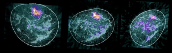
Images of a patient with breast cancer before treatment (left), after one cycle of chemotherapy (center) and just before surgery (right). This patient did not respond well to neoadjuvant chemotherapy and active disease was found in the surgical specimen.
2. High resolution molecular imaging in inflammatory arthritic disease
a. Monitoring of response to targeted therapy in Rheumatoid and Psoriatic Arthritis using high-resolution extremity PET/CT and optical imaging (John C. Hunter, MD, Nancy E. Lane MD, Barton L. Wise, MD, Siba P. Raychaudhuri, MD)
We are developing molecular imaging technologies for (a) early detection of arthritic disease, (b) differential diagnosis of musculoskeletal conditions, and (c) early monitoring of response to treatment in arthritic disease. We are pursuing two imaging approaches – one that uses extremity PET/CT and MRI biomarkers and the other that uses optical imaging biomarkers in conjunction with extremity PET/CT. Recently, the adjoining image was selected as the ‘Image of the month’ by the European Journal of Nuclear Medicine and Molecular Imaging. A free copy of the article is here. Another image from this project will also appear on the cover of the journal Rheumatology for all 12 issues in 2011.
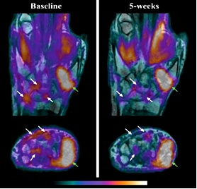
Images show early detection by high resolution PET/MRI of a Rheumatoid Arthritis patient’s response to a TNF-alpha inhibitor. Typically, a standard of care physical exam would have taken 12 weeks.
b. High-resolution PET/CT imaging of the wrist and hand using NaF (Steve Fallen MD, Northern California PET Imaging Center)
This study explores the potential role of high resolution PET/CT imaging using the radioisotope NaF in inflammatory arthritic diseases. The expectation is that osteoblastic activity will be visualized and quantified in more detail using high resolution devices and better assessment of current or novel therapeutic regimens may be possible in the future.
3. PET imaging for Alzheimer’s Disease
Clinical trial of AV-45 for PET imaging for Alzheimer’s Disease (Charles DeCarli, M.D.)
Alzheimer’s disease affects 24 million people worldwide. In its early stages, short-term memory loss is the most common symptom. Later symptoms include confusion, anger, mood swings, language breakdown, long-term memory loss, and the general withdrawal of the sufferer as his or her senses decline. Gradually the sufferer loses minor, and then major bodily functions, until death occurs.
Imaging has played an important role in Alzheimer’s research and is a major topic at UC Davis (http://neuroscience.ucdavis.edu/idealab/). This project aims to test the diagnostic capabilities of a new F-18 labeled PET tracer for amyloid plaques.